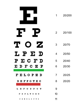
Visual acuity

Visual acuity (VA) commonly refers to the clarity of vision. Visual acuity is dependent on optical and neural factors, i.e., (i) the sharpness of the retinal focus within the eye, (ii) the health and functioning of the retina, and (iii) the sensitivity of the interpretative faculty of the brain.Theodor Wertheim in Berlin presents detailed measurements of acuity in peripheral vision.Hugh Taylor uses these design principles for a 'Tumbling E Chart' for illiterates, later used to study the visual acuity of Australian Aborigines.Rick Ferris et al. of the National Eye Institute chooses the LogMAR chart layout, implemented with Sloan letters, to establish a standardized method of visual acuity measurement for the Early Treatment of Diabetic Retinopathy Study (ETDRS).These charts are used in all subsequent clinical studies, and did much to familiarize the profession with the new layout and progression. Data from the ETDRS were used to select letter combinations that give each line the same average difficulty, without using all letters on each line.The International Council of Ophthalmology approves a new 'Visual Acuity Measurement Standard', also incorporating the above features.Antonio Medina and Bradford Howland of the Massachusetts Institute of Technology develop a novel eye testing chart using letters that become invisible with decreasing acuity, rather than blurred as in standard charts. They demonstrate the arbitrary nature of the Snellen fraction and warn about the accuracy of visual acuity determined by using charts of different letter types, calibrated by Snellen's system.For example, the recording CF 5' would mean the patient was able to count the examiner's fingers from a maximum distance of 5 feet directly in front of the examiner.For example, the recording HM 2' would mean that the patient was able to distinguish movement of the examiner's hand from a maximum distance of 2 feet directly in front of the examiner.A person meets the criteria for permanent blindness under section 95 of the Social Security Act if the corrected visual acuity is less than 6/60 on the Snellen Scale in both eyes or there is a combination of visual defects resulting in the same degree of permanent visual loss.he term 'blindness' means central visual acuity of 20/200 or less in the better eye with the use of a correcting lens. An eye that is accompanied by a limitation in the fields of vision such that the widest diameter of the visual field subtends an angle no greater than 20 degrees shall be considered for purposes in this paragraph as having a central visual acuity of 20/200 or less. Visual acuity (VA) commonly refers to the clarity of vision. Visual acuity is dependent on optical and neural factors, i.e., (i) the sharpness of the retinal focus within the eye, (ii) the health and functioning of the retina, and (iii) the sensitivity of the interpretative faculty of the brain. A common cause of low visual acuity is refractive error (ametropia), or errors in how the light is refracted in the eyeball. Causes of refractive errors include aberrations in the shape of the eyeball or the cornea, and reduced flexibility of the lens. Too high or too low refractive error (in relation to the length of the eyeball) is the cause of nearsightedness (myopia) or farsightedness (hyperopia) (normal refractive status is referred to as emmetropia). Other optical causes are astigmatism or more complex corneal irregularities. These anomalies can mostly be corrected by optical means (such as eyeglasses, contact lenses, laser surgery, etc.). Neural factors that limit acuity are located in the retina or the brain (or the pathway leading there). Examples for the first are a detached retina and macular degeneration, to name just two. Another common impairment, amblyopia, is caused by the visual brain not having developed properly in early childhood. In some cases, low visual acuity is caused by brain damage, such as from traumatic brain injury or stroke. When optical factors are corrected for, acuity can be considered a measure of neural well-functioning. Visual acuity is typically measured while fixating, i.e. as a measure of central (or foveal) vision, for the reason that it is highest there. However, acuity in peripheral vision can be of equal (or sometimes higher) importance in everyday life. Acuity declines towards the periphery in an inverse-linear fashion (i.e. the decline follows a hyperbola). Visual acuity is a measure of the spatial resolution of the visual processing system. VA, as it is sometimes referred to by optical professionals, is tested by requiring the person whose vision is being tested to identify so-called optotypes – stylized letters, Landolt rings, pediatric symbols, symbols for the illiterate, standardized Cyrillic letters in the Golovin–Sivtsev table, or other patterns – on a printed chart (or some other means) from a set viewing distance. Optotypes are represented as black symbols against a white background (i.e. at maximum contrast). The distance between the person's eyes and the testing chart is set so as to approximate 'optical infinity' in the way the lens attempts to focus (far acuity), or at a defined reading distance (near acuity). A reference value above which visual acuity is considered normal is called 6/6 vision, the USC equivalent of which is 20/20 vision: At 6 metres or 20 feet, a human eye with that performance is able to separate contours that are approximately 1.75 mm apart. Vision of 6/12 corresponds to lower, vision of 6/3 to better performance. Normal individuals have an acuity of 6/4 or better (depending on age and other factors). In the expression 6/x vision, the numerator (6) is the distance in metres between the subject and the chart and the denominator (x) the distance at which a person with 6/6 acuity would discern the same optotype. Thus, 6/12 means that a person with 6/6 vision would discern the same optotype from 12 metres away (i.e. at twice the distance). This is equivalent to saying that with 6/12 vision, the person possesses half the spatial resolution and needs twice the size to discern the optotype. A simple and efficient way to state acuity is by solving the fraction to a decimal number. 6/6 then corresponds to an acuity (or a Visus) of 1.0 (see Expression below). 6/3 corresponds to 2.0, which is often attained by well-corrected healthy young subjects with binocular vision. Stating acuity as a decimal number is the standard in European countries, as required by the European norm (EN ISO 8596, previously DIN 58220). The precise distance at which acuity is measured is not important as long as it is sufficiently far away and the size of the optotype on the retina is the same. That size is specified as a visual angle, which is the angle, at the eye, under which the optotype appears. For 6/6 = 1.0 acuity, the size of a letter on the Snellen chart or Landolt C chart is a visual angle of 5 arc minutes (1 arc min = 1/60 of a degree). By the design of a typical optotype (like a Snellen E or a Landolt C), the critical gap that needs to be resolved is 1/5 this value, i.e., 1 arc min. The latter is the value used in the international definition of visual acuity:
